


产品特性
5-HT (Serotonin) Goat Antibody
应用:ICC,IF,IHC,WB验证
Host:Goat;Immunogen:Serotonin
高文献引用数据支持(通过CiteAb官网可查)
| 规格 | 价格 |
|---|---|
| 100 µL | ¥咨询 |
产品咨询:info@biopcr.com
产品订购:sales@biopcr.com
技术支持:tech@biopcr.com
服务热线:400-860-6200
详细介绍
产品名称:5-HT (Serotonin)山羊抗体
别名:anti-5-HT Goat;5-HT Goat Antibody;5-HT抗体;山羊5-羟色胺抗体
宿主:Goat
应用:ICC,IF,IHC,WB验证
免疫原:Serotonin
反应性:Cat, Cod, Cow, Crayfish, Dolphin, Fish, Frog (Xenopus Laevis), Guinea Pig, Hamster, Human, Mexican salamander (Ambystoma Mexicanum), Monkey, Moth, Mouse, Pig, Rabbit, Ram (Sheep), Rat, Salamander, Snail, Tapeworm, Worm, Zebrafish
形式:Lyophilized Whole Serum
描述:
The ImmunoStar serotonin antiserum was quality control tested using standard immunohistochemical methods. The antiserum demonstrates strongly positive labeling of rat hypothalamus and spinal cord using indirect immunofluorescent and biotin/avidin-HRP techniques. Recommended primary dilution is 1/5000 – 1/10,000 in PBS/0.3% Triton X-100 – Bn/Av-HRP Technique.
Staining is completely eliminated by pretreatment of the diluted antibody with 100 µg of serotonin/BSA conjugate.
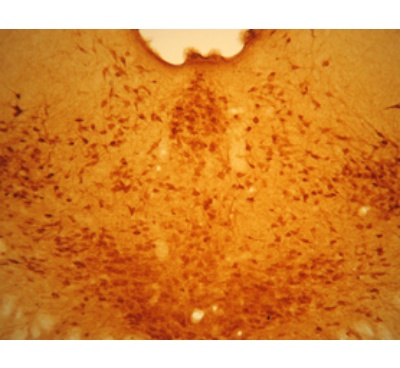
图片说明:
Low magnification of IHC image of neurons staining for the 5-HT goat antibody in the pontine region of the rat brain (top), and high magnification of the raphe nucleus (bottom). The tissue was fixed with 4% formaldehyde/0.05% glutaraldehyde in 0.1 M phosphate buffer, before being removed and prepared for vibratome sectioning. Floating sections were incubated at RT in 10% rabbit serum in PBS, before standard IHC procedure. Primary antibody was incubated at 1:7500 for 48 hours, rabbit anti-goat secondary was subsequently added for 1 hour after washing with PBS. Light microscopy staining was achieved with standard biotin-streptavidin/HRP procedure and DAB chromogen.
RRID:AB_572262
GeneSymbol:Spl
运输条件:Blue ice
返回列表
The ImmunoStar serotonin antiserum was quality control tested using standard immunohistochemical methods. The antiserum demonstrates strongly positive labeling of rat hypothalamus and spinal cord using indirect immunofluorescent and biotin/avidin-HRP techniques. Recommended primary dilution is 1/5000 – 1/10,000 in PBS/0.3% Triton X-100 – Bn/Av-HRP Technique.
Staining is completely eliminated by pretreatment of the diluted antibody with 100 µg of serotonin/BSA conjugate.
图片说明:
Low magnification of IHC image of neurons staining for the 5-HT goat antibody in the pontine region of the rat brain (top), and high magnification of the raphe nucleus (bottom). The tissue was fixed with 4% formaldehyde/0.05% glutaraldehyde in 0.1 M phosphate buffer, before being removed and prepared for vibratome sectioning. Floating sections were incubated at RT in 10% rabbit serum in PBS, before standard IHC procedure. Primary antibody was incubated at 1:7500 for 48 hours, rabbit anti-goat secondary was subsequently added for 1 hour after washing with PBS. Light microscopy staining was achieved with standard biotin-streptavidin/HRP procedure and DAB chromogen.
RRID:AB_572262
GeneSymbol:Spl
运输条件:Blue ice