


产品特性
Neuropeptide Y Y1 Receptor Antibody
应用:ICC,IF,IHC,WB验证
Host:Rabbit
高文献引用数据支持(通过CiteAb官网可查)
| 规格 | 价格 |
|---|---|
| 100 µL | ¥咨询 |
产品咨询:info@biopcr.com
产品订购:sales@biopcr.com
技术支持:tech@biopcr.com
服务热线:400-860-6200
详细介绍
产品名称:Neuropeptide Y Y1 Receptor抗体
别名:Neuropeptide Y receptor 1; NPY receptor 1; NPYR; NPYY1; NPY1-R; Neuropeptide Y receptor type 1; FC5; NPYR1, neuropeptide Y1 receptor, anti-NPYY1
宿主:Rabbit
应用:ICC,IF,IHC,WB验证
反应性:Hamster, Human, Mouse, Rat
形式:液体
描述:
The ImmunoStar Neuropeptide Y Y1 Receptor was quality control tested using standard immunohistochemical methods. The antiserum demonstrates strongly positive labeling of rat cortex, arcuate and hippocampus using indirect immunofluorescent and biotin/avidin-HRP techniques. Recommended primary dilutions are 1/500 – 1/1000 in PBS – Bn/Av-HRP detection.
The three black and white images are confocal image of spinal cord neurons labeled with antibody 24506 against the neuropeptide Y Y1 receptor using immunofluorescence. More details in Marvizon et al., 2019, Neuropharmacology 158: 107732. Courtesy of Juan Carlos Marvizon, Ph.D., UCLA.
The antibody was characterized by immunohistochemistry and Western blot. Western blot showed one immunoreactive band of 40 kD and a single high molecular weight band, presumably a precursor molecule. Due to the difficulty with receptor antibodies, western blot applications are not warranted and are included as specificity information only.
Preincubation of the antibody with an excess of the synthetic peptide blocked staining. Immunohistochemical staining of rat brain correlates well with Northern analysis, in situ hybridization and receptor autoradiography. BlastP database sequence homology searches confirmed that this sequence is unique to rat, mouse and human NPY Y1 receptors.
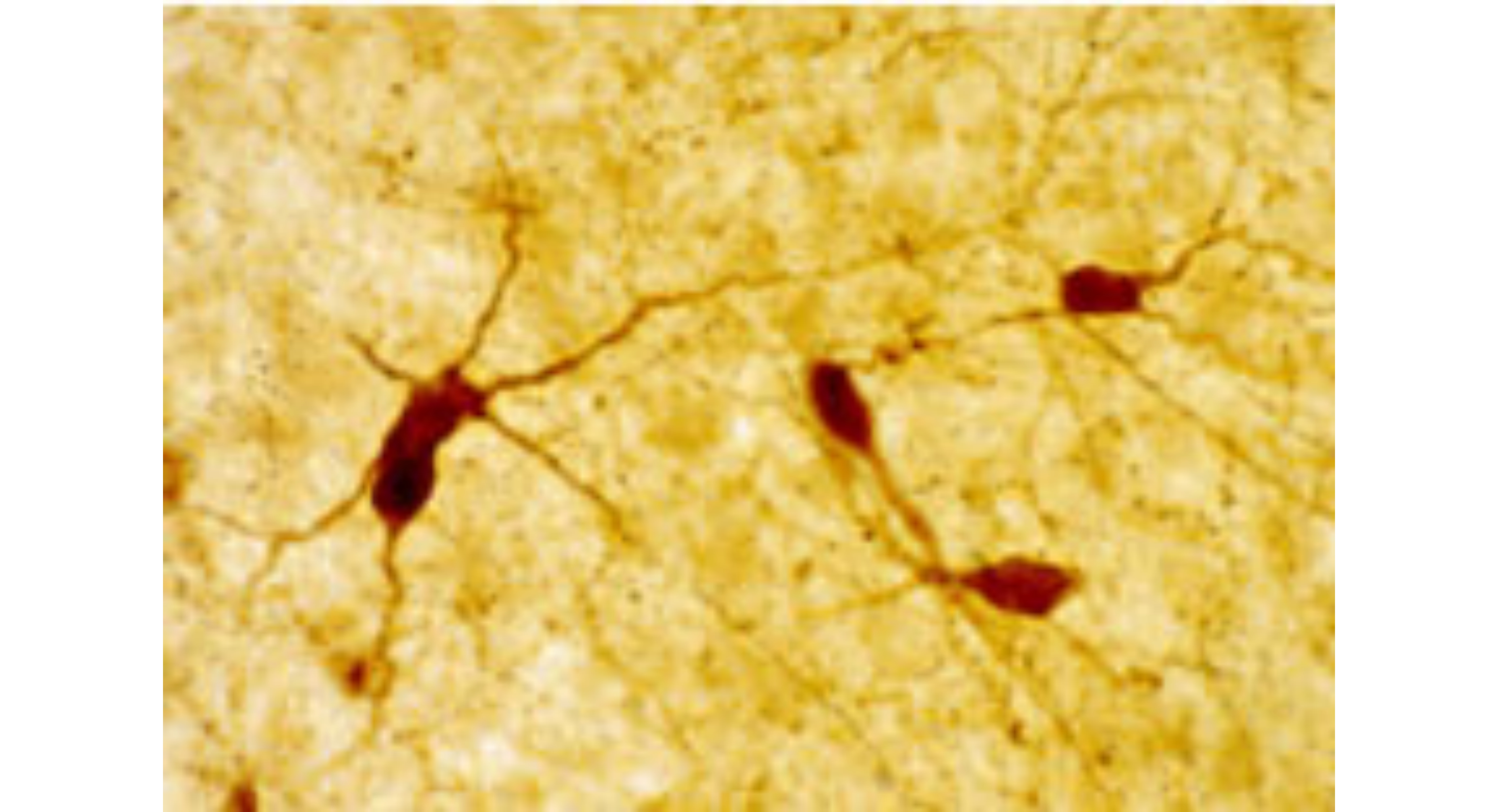
图片说明:
IHC image of the rat cortex staining for the neuropeptide Y Y1 recptor. The tissue was fixed with 4% formaldehyde/0.05% glutaraldehyde in 0.1 M phosphate buffer, before being removed and prepared for vibratome sectioning. Floating sections were incubated at RT in 10% goat serum in PBS, before standard IHC procedure. Primary antibody was incubated at 1:5000 for 48 hours, goat anti-rabbit secondary was subsequently added for 1 hour after washing with PBS. Light microscopy staining was achieved with standard biotin-streptavidin/HRP procedure and DAB chromogen.
GeneSymbol:neuropeptideYreceptorY1,anti-NPYY1
运输条件:Blue ice
返回列表
The ImmunoStar Neuropeptide Y Y1 Receptor was quality control tested using standard immunohistochemical methods. The antiserum demonstrates strongly positive labeling of rat cortex, arcuate and hippocampus using indirect immunofluorescent and biotin/avidin-HRP techniques. Recommended primary dilutions are 1/500 – 1/1000 in PBS – Bn/Av-HRP detection.
The three black and white images are confocal image of spinal cord neurons labeled with antibody 24506 against the neuropeptide Y Y1 receptor using immunofluorescence. More details in Marvizon et al., 2019, Neuropharmacology 158: 107732. Courtesy of Juan Carlos Marvizon, Ph.D., UCLA.
The antibody was characterized by immunohistochemistry and Western blot. Western blot showed one immunoreactive band of 40 kD and a single high molecular weight band, presumably a precursor molecule. Due to the difficulty with receptor antibodies, western blot applications are not warranted and are included as specificity information only.
Preincubation of the antibody with an excess of the synthetic peptide blocked staining. Immunohistochemical staining of rat brain correlates well with Northern analysis, in situ hybridization and receptor autoradiography. BlastP database sequence homology searches confirmed that this sequence is unique to rat, mouse and human NPY Y1 receptors.
图片说明:
IHC image of the rat cortex staining for the neuropeptide Y Y1 recptor. The tissue was fixed with 4% formaldehyde/0.05% glutaraldehyde in 0.1 M phosphate buffer, before being removed and prepared for vibratome sectioning. Floating sections were incubated at RT in 10% goat serum in PBS, before standard IHC procedure. Primary antibody was incubated at 1:5000 for 48 hours, goat anti-rabbit secondary was subsequently added for 1 hour after washing with PBS. Light microscopy staining was achieved with standard biotin-streptavidin/HRP procedure and DAB chromogen.
GeneSymbol:neuropeptideYreceptorY1,anti-NPYY1
运输条件:Blue ice